The Biology Behind Long-Term Implant Success
September 25, 2026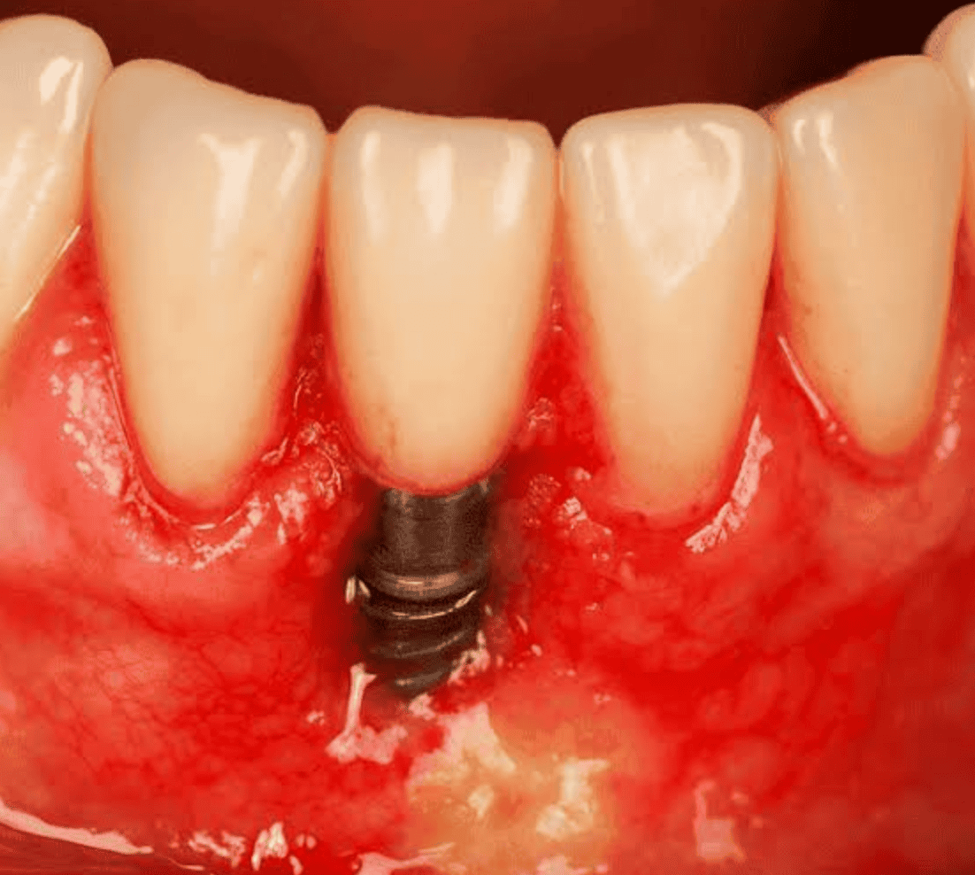
As dental implant procedures become increasingly routine, clinicians face a growing secondary challenge: managing peri-implant disease. Among these conditions, peri-implantitis characterized by inflammation of the peri-implant mucosa and progressive loss of supporting marginal bone poses a significant threat to long-term implant survival.
While much of the clinical focus on peri-implantitis centers on treatment, the reality is that therapeutic options can be unpredictable and complex. Effective management hinges on a simple truth: prevention begins long before the surgical site is prepared.
1. Pre-Operative Risk Assessment: Identifying Vulnerabilities Early
Preventing peri-implantitis starts during the initial patient consultation. Identifying patient-specific risk factors allows clinicians to modify treatment plans, optimize systemic health, and establish appropriate maintenance schedules before placing a single fixture.
- History of Periodontitis: Patients with a history of treated or active periodontitis face a significantly higher risk of peri-implantitis. The same pathogenic microflora responsible for periodontal tissue destruction can colonize peri-implant pockets.
- Smoking and Nicotine Use: Nicotine impairs microvascular perfusion, alters immune response, and compromises osseointegration and soft-tissue healing.
- Systemic Conditions (e.g., Uncontrolled Diabetes): Hyperglycemia impairs neutrophil function and wound healing, predisposing tissues to persistent inflammation and bone loss.
- Inadequate Keratinized Tissue: A narrow band or absence of keratinized mucosa (<2 mm) around an implant makes plaque control uncomfortable for the patient and renders the soft tissue seal more vulnerable to mechanical trauma.
- Poor Oral Hygiene: Plaque accumulation is the primary driver of peri-implant mucositis, the reversible precursor to peri-implantitis.
2. Surgical & Prosthetic Factors: Designing for Prevention
Proper planning and execution minimize the risk of biological complications. Technical errors during the surgical or restorative phases frequently contribute to peri-implant tissue breakdown.
- Ideal 3D Implant Placement: Implants placed too far buccally, too close together (<3 mm between implants), or too close to adjacent teeth (<1.5 mm) lead to compromised bone thickness and soft tissue contours that are difficult for patients to clean.
- Excess Cement Retention: Subgingival residual cement is one of the most common, preventable triggers for peri-implantitis. Utilizing screw-retained restorations or custom abutments with margin placements near the gingival peak significantly reduces this risk.
- Prosthetic Emergence Profile: Oversized or convex emergence profiles hinder oral hygiene access and compress surrounding soft tissue, creating stagnation areas for plaque accumulation.
- Surgical Trauma: Overheating bone during osteotomy preparation or failing to achieve primary stability can lead to early marginal bone loss, predisposing the site to future infection.
3. Early Diagnosis: What to Look For
Early detection is essential for arresting disease progression. Regular monitoring allows clinicians to differentiate between healthy peri-implant tissue, peri-implant mucositis, and peri-implantitis.
Diagnostic Criteria for Peri-Implantitis
- Bleeding on Probing (BOP) and/or Suppuration: Visual signs of inflammation combined with gentle probing (approx. 0.25 N) indicating micro-ulceration of the pocket epithelium.
- Increased Probing Depths: A baseline probing depth should be recorded at the time of final prosthesis insertion. Increases in depth over time signal tissue breakdown.
- Progressive Bone Loss: Radiographic evidence of marginal bone loss beyond initial peri-implant remodeling (typically >2 mm from the baseline post-loading radiograph).
4. Maintenance and Prevention Protocols
Long-term success relies on a structured supportive peri-implant therapy (SPIT) program combined with effective patient self-care.
In-Office Maintenance
- Routine Monitoring: Record probing depths, BOP, and suppuration at every recall visit. Take baseline radiographs post-restoration and update them periodically based on clinical risk.
- Professional Debridement: Utilize non-abrasive instruments—such as titanium cuvettes, carbon-fiber scalers, or specialized air-polishing systems with glycine/erythritol powder—to clean implant surfaces without scratching the titanium surface.
Patient Home Care
- Tailored Oral Hygiene Aids: Recommend interdental brushes (with plastic-coated wires), specialized dental floss, or water flossers to clean hard-to-reach prosthetic contours.
- Antimicrobial Adjuncts: Short-term use of chlorhexidine rinses or daily application of targeted antimicrobial gels can help manage localized inflammation during flare-ups.
Summary
Preventing peri-implantitis requires a proactive approach rather than a reactive one. By evaluating risk factors prior to surgery, ensuring precise 3D placement, designing hygienic restorations, and establishing an unyielding supportive maintenance protocol, clinicians can safeguard both the implant and the patient’s long-term oral health.